|
Adminஐﻬ
|
 |
« on: December 17, 2005, 06:17:17 pm » |
|
Lupus and Scleroderma Lupus 
The variety of skin rashes seen in lupus are due to inflammation, rather than fibrosis. Features of these rashes are limited to the skin surfaces exposed. They include:
- the facial "butterfly" rash
- a photosensitivity reaction (rash, hives or blisters) seen immediately after exposure to sunlight or other sources of ultraviolet light.
An exception is "discoid" or "cutaneous" lupus, which consists of spots or patches of rash, mostly in sun-exposed areas (face, ears, extremities), which typically cause scarring and skin pigment changes. The appearance of scleroderma and discoid lupus are entirely different, and should be easily distinguished from one another by your physician.
Scleroderma
The hallmark of scleroderma (SSc) is thickened skin (sclero=hard; derma=skin). If skin thickening is widespread, it may extend to the upper arms, thighs, chest, and abdomen.
These changes are due to the excessive production and uncontrolled "laydown" of collagen, the substance normally present in scar tissue.
If skin fibrosis (hardening) is widespread, it may extend to:
- the upper arms
- thighs
- chest
- abdomen.
The variety of skin rashes seen in lupus are due to inflammation, rather than fibrosis. These include the facial butterfly rash and photosensitivity reaction (rash, hives or blisters ) seen immediately after exposure to sunlight or other sources of ultraviolet light ). The latter is limited to the skin surfaces exposed. An exception is discoid lupus , which consists of spots or patches of rash, mostly in sun exposed areas (face, ears, extremities), which typically cause scarring and skin pigment changes. The appearance of scleroderma and discoid lupus are entirely different, and should be easily distinguished from one another by your physician.
Other features less common in lupus than in SSc include:
- pulmonary fibrosis: scarring of the lower portions of the lung
- difficulty in swallowing solid foods such as bread or meat
- heartburn or indigestion from stomach acid "refluxing" ( coming back) into the esophagus. Difficulty swallowing and reflux are due to sluggish and uncoordinated motion of the muscle layer of the esophagus (esophageal dysmotility).
-Scleroderma often leads to finger and hand deformities as well, due to the combination of skin thickening, arthritis, tendinitis and tenosynovitis (inflammation and scarring of tendons and their lining tissues). These processes ultimately result in limited movement of the fingers-Raynaud's phenomenon: fingers turn blue or white with cold. This occurs in 95 percent of persons with scleroderma and in 40 percent of persons with lupus.
Treatment
The primary treatment approaches to SSc are quite different from those for lupus.
Therefore, treatment for scleroderma like problems in persons with lupus should be individualized and directed at the particular problems present at any given time. |
|
|
|
« Last Edit: June 06, 2007, 03:27:27 pm by Admin »
|
 Logged
|
 I look normal, as I have an "Invisible Illness". You can not catch it, you can not see it. It's called Lupus.My body is attacking itself on the inside.  www.LupusMCTD.com www.LupusMCTD.com Represents: 1) We are patients helping researchers build a future for the lives of others... 2) Where HOPE is a WORK In Progress 3) Pay It Forward~Giving Back To The Future Lupus/MCTD Patients |
|
|
|
Adminஐﻬ
|
|
« Reply #1 on: December 21, 2005, 07:09:24 pm » |
|
|
|
|
|
« Last Edit: June 06, 2007, 03:25:41 pm by Admin »
|
Logged
|
I look normal, as I have an "Invisible Illness". You can not catch it, you can not see it. It's called Lupus.My body is attacking itself on the inside. www.LupusMCTD.com Represents: 1) We are patients helping researchers build a future for the lives of others... 2) Where HOPE is a WORK In Progress 3) Pay It Forward~Giving Back To The Future Lupus/MCTD Patients |
|
|
|
Adminஐﻬ
|
|
« Reply #2 on: December 24, 2005, 12:05:02 pm » |
|
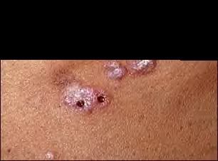 Lupus, discoid - view of lesions on the chest: This close-up picture of the neck clearly shows the typical rounded appearance of discoid lupus. The whitish appearance is caused by scaling. The two dark spots are biopsy sites and are not part of the disease. *NOTE this picture file is too large that is why you see the black area.. But this gives you a general idea of Discoid Lupus looks like. Once I find a smaller size file pic, I'll replace it. But I liked this one, it shows great detail, plus the punched biopsy to also give you an idea how big/small it is, in the event you need to have one done. ~~~~~~~~~~~~~~~~~~~~~~~~~~~~~~~~~~~~~~~~~~~~~~~~~~~~~~~~~~~ Re: Child photo below: The rash of lupus is round or disk shaped (discoid) and is characterized by red, raised patches with adherent scales. The skin pores (follicles) may be plugged, and scarring often occurs in older lesions. Approximately 90% of individuals with discoid lupus have only skin involvement as compared to more generalized involvement in systemic lupus erythematosis (SLE).[/size] |
|
|
|
« Last Edit: May 17, 2006, 06:33:06 pm by Kathy »
|
Logged
|
I look normal, as I have an "Invisible Illness". You can not catch it, you can not see it. It's called Lupus.My body is attacking itself on the inside. www.LupusMCTD.com Represents: 1) We are patients helping researchers build a future for the lives of others... 2) Where HOPE is a WORK In Progress 3) Pay It Forward~Giving Back To The Future Lupus/MCTD Patients |
|
|
|
Adminஐﻬ
|
|
« Reply #3 on: January 17, 2006, 09:16:01 pm » |
|
Dermatomyositis
Overview
Dermatomyositis (dur-muh-to-mi-uh-SI-tis) is an uncommon disease marked by muscle weakness and a distinctive skin rash. Because of similarities in signs, symptoms and treatment, dermatomyositis is often discussed in conjunction with polymyositis.
Both conditions fall into the category of inflammatory muscle diseases "myo" means "muscles" in Greek; "itis" means "inflamed." "Derma," which means "skin," implies the skin-related signs and symptoms that accompany the muscle inflammation of dermatomyositis.
Although dermatomyositis may occur at any age, it mostly affects adults in their late 40s to early 60s or children between 5 and 15 years of age. Women have dermatomyositis more often than men do. Dermatomyositis in children is distinct from the adult form. The disease usually develops over weeks or months.
Periods of remission, during which signs and symptoms of dermatomyositis improve spontaneously, may occur. Treatment can improve your skin and your muscle strength and function.
Signs and symptoms
The most common signs and symptoms of dermatomyositis include:
A violet-colored or dusky red rash, most commonly on your face, eyelids, and areas around your nails, knuckles, elbows, knees, chest and back. Affected areas are typically more sensitive to sun exposure.
Progressive muscle weakness, particularly in the muscles closest to the trunk, such as those in your hips, thighs, shoulders, upper arms and neck. This weakness is symmetrical, affecting both the left and right sides of your body.
Difficulty swallowing (dysphagia).
Muscle pain or tenderness.
Fatigue, fever and weight loss.
Hardened deposits of calcium under the skin (calcinosis), especially in children.
Gastrointestinal ulcers and infections, also more common in children.
Lung problems.
The skin rash usually occurs at the same time as muscle weakness, but may precede muscle weakness by a few weeks. Sometimes, the skin rash alone determines the diagnosis. In some children with dermatomyositis, the skin may become thick and hard in a way similar to scleroderma. When this happens, the condition is called sclerodermatomyositis.
Weakness in muscles, such as your hips and shoulders, can lead to difficulty in getting out of chairs, climbing stairs, brushing your hair or working with your arms over your head. Weakness in your neck muscles can make it hard to hold your head up.
Causes
Dermatomyositis belongs to a group of conditions called inflammatory myopathies. Myopathies are diseases or abnormal conditions of the striated muscles that cover your skeleton. The cause of most inflammatory myopathies is unknown. Other inflammatory muscle diseases include inclusion body myositis, which progresses more slowly than other forms; myositis associated with other connective tissue diseases, such as lupus or scleroderma; and myositis associated with cancer (malignancy).
Doctors suspect that inflammatory myopathies are autoimmune disorders, in which your immune system turns against normal body components. Infections caused by bacteria, parasites or viruses can cause inflammatory myopathies, but in most cases, doctors aren't able to identify a preceding infection in dermatomyositis. Some doctors think certain people may have a genetic susceptibility to the disease.
Typically, your immune system works to protect your healthy cells from attacks by foreign substances, such as bacteria and viruses. If you have dermatomyositis, an unknown cause seems to trigger your immune system to begin producing autoimmune antibodies (also called autoantibodies) that attack your body's own tissues. Small blood vessels in muscular tissue appear to be particularly affected. Destruction of these blood vessels eventually leads to degeneration of muscle fibers. Many people with dermatomyositis show a detectable level of autoantibodies in their blood. It's unclear whether these autoantibodies are involved in causing dermatomyositis.
When to seek medical advice
If you experience any of the signs or symptoms of dermatomyositis, see your doctor. The earlier the disease is detected, the better your response may be to treatment. With treatment, you can manage and sometimes even reverse your signs and symptoms.
If you have difficulty swallowing or shortness of breath, call your doctor right away, as this may indicate rapid progression of the disease and the need for immediate help.
Screening and diagnosis
Dermatomyositis is the most easily recognized of the inflammatory muscle diseases because of its characteristic rash. Occasionally, a rash alone may prompt a diagnosis of dermatomyositis without muscle involvement (amyopathic dermatomyositis).
In addition to a thorough evaluation, including examination of muscle strength and a detailed family medical history, your doctor may use some or all of the following tests to assist in the diagnosis:
Electromyography. A doctor with specialized training inserts a thin needle electrode through the skin into the muscle to be tested. Electrical activity is measured as you relax or tighten the muscle. Changes in the pattern of electrical activity can confirm a muscle disease. The doctor can determine the distribution of the disease by testing different muscles.
Blood analysis. A blood test will let your doctor know if you have elevated levels of muscle enzymes, such as creatine kinase (CK) and aldolase. Increased CK and aldolase levels can indicate muscle damage. A blood test can also determine whether autoantibodies are present in your blood.
Muscle biopsy. A small piece of muscle tissue is removed surgically for laboratory analysis. A muscle biopsy may reveal abnormalities in your muscles, such as inflammation, damage or infection. The tissue sample can also be examined for the presence of abnormal proteins and checked for enzyme deficiencies.
Magnetic resonance imaging (MRI). A scanner creates cross-sectional images of your muscles from data generated by a powerful magnetic field and radio waves. These images can be viewed from any direction or plane. During the test, you lie inside the cylindrical-shaped scanner for 15 minutes to an hour. MRI scans may help detect inflammation in your muscles.
Complications
If the muscles in your esophagus are affected, you may have problems swallowing (dysphagia), which in turn may cause weight loss and malnutrition. Dysphagia may also lead to entrance of food or liquids, including saliva, into your lungs (aspiration), which can lead to pneumonia. If your chest muscles are involved, you may experience breathing problems, such as shortness of breath. Gastrointestinal ulceration and bleeding can occur. Deposits of calcium in your muscles, skin and connective tissues (calcinosis) can occur late in the disease, particularly if you've had the disease for a long time.
Dermatomyositis may be associated with other conditions, including:
Raynaud's phenomenon. This is a condition in which your fingers, toes, cheeks, nose and ears turn pale when exposed to cold temperatures.
Other connective tissue diseases. Diseases that affect tissues that hold your body together, such as your muscles and joints, sometimes occur in conjunction with each other. Conditions such as lupus, rheumatoid arthritis, scleroderma and Sjogren's syndrome can occur in combination with dermatomyositis.
Cardiovascular disease. The muscle of your heart may become inflamed (myocarditis). In a small number of people who have dermatomyositis, congestive heart failure and heart arrhythmias may develop.
Lung disease. A condition called interstitial lung disease may occur with dermatomyositis. Interstitial lung disease refers to a group of disorders that cause scarring (fibrosis) of lung tissue, making lungs stiff and inelastic. Signs and symptoms include a dry cough and shortness of breath.
Cancer. Dermatomyositis in adults has been linked to an increased likelihood of cancer, particularly of the lungs, breasts, ovaries and gastrointestinal tract. Risk of cancer increases with age, although it appears to level off three years after a diagnosis of dermatomyositis.
The outcome (prognosis) of dermatomyositis tends to be better if there are no associated complications, such as esophageal damage, lung disease or cancer. Early diagnosis is important, because it can lead to prompt treatment and evaluation of associated conditions. Early treatment of children can prevent the development of calcinosis, a condition that can be hard to treat once it's established.
Pregnancy may worsen signs and symptoms in women with active disease. Active dermatomyositis can also increase the risk of premature birth or stillbirth. If the disease is in remission, the risk isn't as great.
People with polymyositis or dermatomyositis may also be at an increased risk of infections, particularly respiratory and digestive infections. As a result, your doctor may monitor your signs and symptoms for any indication of infection so that you can receive prompt diagnosis and treatment.
Treatment
Although there's no cure for dermatomyositis, treatment can improve your skin, muscle strength and function. Treatment begun early in the disease process tends to be more effective, often because there are fewer complications. Methods of therapy include the following:
Corticosteroids. These medications suppress your immune system, limiting the production of antibodies and reducing skin and muscle inflammation. Corticosteroids, especially prednisone, are usually the first choice in treating inflammatory myopathies such as dermatomyositis. Your doctor may start with a very high dose, and then decrease it as your signs and symptoms improve. This generally takes about two to four weeks. Your doctor may also prescribe topical corticosteroids for your skin. Visible results are usually evident within three to six months, but therapy is often needed for years. Prolonged use of corticosteroids can have serious side effects including osteoporosis, weight gain, diabetes, increased risk of someinfections, mood swings, cataracts, high blood pressure, a redistribution of body fat and muscle weakness. As a result, your doctor may also recommend supplements such as calcium and vitamin D and may prescribe bisphosphonates such as alendronate (Fosamax) or risedronate (Actonel).
Immunosuppressants. If your body doesn't respond adequately to corticosteroids, your doctor may recommend other immunosuppressive drugs such as azathioprine (Imuran) or methotrexate (Rheumatrex). Your doctor may prescribe these alone or in combination with corticosteroids. When in combination, these additional immunosuppressants can be used to lessen the dose and potential side effects of the corticosteroid. Immunosuppressants such as cyclophosphamide (Cytoxan) and cyclosporine (Neoral, Sandimmune) may improve signs and symptoms of dermatomyositis and interstitial lung disease.
Antimalarial medications. For a persistent rash, your doctor may prescribe an antimalarial medication such as hydroxychloroquine (Plaquenil) or chloroquine phosphate (Aralen). Be aware, however, that adverse reactions to such medications, though uncommon, can be confused with the rash or weakness of dermatomyositis itself.
Intravenous immunoglobulin (IVIg). This involves receiving intravenous infusions of antibodies from a group of donors over two to five days. This treatment is still under investigation and is usually expensive. It may be an option for you if your dermatomyositis is severe or resistant to other forms of therapy.
Physical therapy. A physical therapist can show you various exercises to maintain and improve your strength and flexibility and advise an appropriate level of activity. Your exercise program is likely to change during the course of the disease and treatment period. Keeping active in general and pacing yourself will help maintain muscle strength.
Surgery. Surgery may be an option to remove painful calcium deposits.
Pain relievers. Over-the-counter drugs such as aspirin, ibuprofen (Advil, Motrin, others) and acetaminophen (Tylenol, others), can be used to treat any accompanying pain. If these aren't sufficient, your doctor may prescribe a stronger pain reliever, such as codeine.
Treatments with unknown long-term effects include:
Plasmapheresis. This treatment, also called plasma exchange, is a type of blood cleansing in which damaging antibodies are removed from your blood.
Radiation therapy. This involves irradiation of the lymph nodes to suppress your immune system.
Treatments that are still in the experimental phase but have shown signs of being effective include fludarabine (Fludara), an agent that prevents the development and growth of malignant cells, and tacrolimus (Prograf), a transplant-rejection drug that may work to inhibit the immune system. Tacrolimus is often used topically to treat dermatomyositis and other skin problems.
Coping skills
Living with a chronic autoimmune disease can make you wonder at times whether you're up to the challenge. Though it isn't always easy, you're tougher than you may think. To help you cope, try supplementing your medical care with the following suggestions:
Know your illness. Read all you can about dermatomyositis and other muscle and autoimmune disorders. Talk to other people who have a similar condition. Don't be afraid to ask your doctor any questions that you may have concerning your illness, diagnosis or treatment plan.
Be a part of your medical team. Consider yourself, your doctor and any other medical experts involved as a united front in the fight against your disease. Following the treatment plan you agreed to is vital. Keep your doctor updated on any new signs or symptoms you may experience. Regularly perform the physical exercises prescribed for you. A speech therapist can help you with swallowing difficulties and a registered dietitian can help you with preparing easy-to-eat foods.
Treat yourself well. Don't wait until you're exhausted to rest. This will only set you back further as your body tries to recuperate. Learning to pace yourself can help you maintain a consistent level of energy, accomplish just as much and feel better emotionally. Learn to say no effectively and ask for help when you need it. Take hot baths, stretch, exercise.
Wear sunscreen. Areas affected by your rash are more sensitive to the sun, so wear protective clothing or high-protection sunscreen when you go out.
Use electric appliances, power tools and other aids. Save your energy using power appliances, such as battery-operated toothbrushes, electric can openers and power screwdrivers. If your ability to walk is impaired, consider using an aid such as a cane, walker or wheelchair.
Accept your emotions. Denial, anger and frustration are normal feelings when you must deal with an illness. Things don't seem normal or fair and likely seem out of your control. Feelings of fear and isolation are common, so stay close to your family and friends. Try to maintain your daily routine as best you can and don't neglect doing those things you enjoy. Many people find support groups to be a helpful resource
This website is provided for educational and informational purposes only. Lupus and Tissue Disorder is not engaged in rendering medical advice or professional services and this information should not be used for diagnosing or treating a health problem. The site author and the content providers make no representation or warranties, expressed or implied. Providing links to other websites does not imply that Lupus and Connective Tissue Disorder endorses the information or services provided on those websites. The organizations operating those websites are solely responsible for the content found on their websites. All material on this web site is copyright © 2005 by Lupus and Mixed Connective Tissue Disorder.
|
|
|
|
« Last Edit: June 06, 2007, 03:29:26 pm by Admin »
|
Logged
|
I look normal, as I have an "Invisible Illness". You can not catch it, you can not see it. It's called Lupus.My body is attacking itself on the inside. www.LupusMCTD.com Represents: 1) We are patients helping researchers build a future for the lives of others... 2) Where HOPE is a WORK In Progress 3) Pay It Forward~Giving Back To The Future Lupus/MCTD Patients |
|
|
|
Adminஐﻬ
|
|
« Reply #4 on: April 12, 2006, 06:10:55 pm » |
|
 Definition: Definition:
Discoid lupus erythematosus is a chronic skin condition characterized by inflammation and scarring type skin lesions which occur on the face, ears, scalp and at times on other body areas. These lesions develop as an inflamed growth with, scaling and a warty like appearance. The center areas may appear lighter in color surrounded by an area darker than the normal skin. When lesions occur in hairy areas such as the scalp, permanent scarring and hair loss can occur. A small percentage of patients with discoid lupus can develop disease of the internal organs which can make the person sick.
II. Workup:
If your doctor suspects that you have this condition, a skin biopsy needs to be done to confirm the diagnosis because other conditions can look like discoid lupus erythematosus.
If the skin biopsy shows discoid lupus erythematosus, then further testing may be indicated.
III. Causes:
The cause is unknown.
This condition tends to run in families.
Females outnumber males with this condition 3 to 1.
In some patients with discoid lupus erythematosus, sunlight may make the lesions come out.
IV. Course:
The lesions can cause permanent scarring and hair loss. The disease usually comes and goes. Discoid lupus will rarely go away and not come back.
V. Treatment:
Cortisone ointment applied to the skin in the involved areas will often improve the lesions and slow down their progression.
Cortisone injections into the lesions will also treat discoid lupus and usually are more effective than the ointment form of cortisone.
If the lesions are becoming unsightly and you really feel something more needs to be done, a drug named Plaquenil will often improve the condition. Patients on Plaquenil need eye exams once a year to prevent damage to the retina of the eye and periodic blood work.
Patients whose condition is sensitive to sunlight need to wear a sunscreen of SPF 15 or higher daily, and a hat while out doors.
Follow-up with the doctor is important and necessary every six months to once a year to make sure the disease is not spreading to the internal organs and to minimize scarring. |
|
|
|
« Last Edit: May 17, 2006, 05:32:58 pm by Kathy »
|
Logged
|
I look normal, as I have an "Invisible Illness". You can not catch it, you can not see it. It's called Lupus.My body is attacking itself on the inside. www.LupusMCTD.com Represents: 1) We are patients helping researchers build a future for the lives of others... 2) Where HOPE is a WORK In Progress 3) Pay It Forward~Giving Back To The Future Lupus/MCTD Patients |
|
|
|
Adminஐﻬ
|
|
« Reply #5 on: May 08, 2006, 08:01:38 pm » |
|
Vasculitis
Inflammation of blood vessels, which is often segmental, may be generalized or localized and constitutes the basic pathogenetic process of various rheumatic diseases and syndromes.
Pathology
Vasculitis may follow various etiologic mechanisms, but the histologic abnormalities are limited. In acute lesions, the predominant inflammatory cells are PMNs; in chronic lesions, lymphocytes. The inflammation is often segmental, with scattered foci of intense inflammation in an otherwise normal vascular tree. Variable degrees of cellular infiltration and necrosis or scarring within one or more layers of the vessel wall occur at the affected sites. Inflammation within the media of a muscular artery tends to destroy the internal elastic lamina. Inflammation at any point in the vessel wall tends to resolve by fibrosis and intimal hypertrophy. Distinguishing histologic features are occasionally seen (eg, numerous giant cells, patchy areas of fibrinoid necrosis where complete sections of the vessel wall have undergone inflammatory destruction and liquefaction). Secondary occlusion of the lumen resulting from intimal hypertrophy or intraluminal thrombus formation is frequent. In addition, once the vessel wall integrity is breached, RBCs and fibrin may leak into the surrounding connective tissue.
Any type and size of vessel may be involved--arteries, arterioles, veins, venules, or capillaries. However, most of the pathophysiology can be ascribed to arterial inflammation, with the potential for partial or total vascular occlusion and subsequent tissue necrosis. Because vasculitis is often segmental or focal, biopsy of clinically suspicious tissue may not always provide definitive histologic diagnosis. However, the intimal and periadventitial fibrous response to a focus of intense vessel wall inflammation frequently extends up and down the vessel from the primary insult, so intimal hypertrophy and fibrosis or perivasculitis on histology suggest the presence of an adjacent area of vasculitis.
Classification
The numerous vasculitic disorders are most usefully classified according to the size and depth of the predominant vessel involved (see also individual discussions elsewhere in The Manual). The depth of lesions is determined by working viscerally from the integument. Table 50-5 lists some of the vasculitic disorders according to the type of inflammation and the clinical presentation.
Many other diseases are also characterized by or strongly associated with vasculitis. Rheumatoid nail-fold infarcts, leg ulcers, and other lesions of the rheumatic diseases appear to have central foci of vasculitis as their basic pathogenesis. Much of SLE pathophysiology can be ascribed to vasculitis with or without secondary vascular occlusion, which is particularly evident in the renal glomerular tufts. Polymyositis or dermatomyositis of childhood frequently includes an element of vasculitis in muscle and at extramuscular and extracutaneous sites. Even the bland-appearing and extensive intimal proliferation of small arteries, which typifies systemic sclerosis, may be a postinflammatory event. Other syndromes dominated by serious vasculitis include polyarteritis nodosa and Wegener's granulomatosis.
|
|
|
|
« Last Edit: May 17, 2006, 04:47:37 pm by Kathy »
|
Logged
|
I look normal, as I have an "Invisible Illness". You can not catch it, you can not see it. It's called Lupus.My body is attacking itself on the inside. www.LupusMCTD.com Represents: 1) We are patients helping researchers build a future for the lives of others... 2) Where HOPE is a WORK In Progress 3) Pay It Forward~Giving Back To The Future Lupus/MCTD Patients |
|
|
|
Adminஐﻬ
|
|
« Reply #6 on: May 29, 2006, 02:30:45 pm » |
|
Lupus Vasculitis Underlies Skin Denervation
To better understand why some lupus patients develop a loss of nerves in the skin, the researchers of this study measured density of nerve fibers within the skin and looked at the relationship of nerve density to lupus activity, the sensitivity of nerves to touch, and the electric activity associated with the use of different parts of the body. The authors concluded that blood vessel inflammation in the skin (cutaneous vasculitis) may lead to the loss of nerves in the skin in lupus patients, and this might lead to dysfunction in the perception of heat and cold
ABSTRACT:
Skin denervation and cutaneous vasculitis in systemic lupus erythematosus
Ming-Tsung Tseng1,4, Song-Chou Hsieh2, Chia-Tung Shun3,5, Kuang-Lun Lee2, Chun-Liang Pan1,8, Whei-Min Lin6, Yea-Hui Lin1, Chia-Li Yu2,7 and Sung-Tsang Hsieh1,6
Departments of 1 Neurology, 2 Internal Medicine and 3 Pathology, National Taiwan University Hospital, 4 Section of Neurology, Department of Internal Medicine, Far Eastern Memorial Hospital, Departments of 5 Forensic Medicine and 6 Anatomy and Cell Biology and 7 Graduate Institute of Molecular Medicine, National Taiwan University College of Medicine, Taipei, Taiwan and 8 Helen Wills Neuroscience Institute, University of California, Berkeley, CA, USA
Correspondence to: Dr Sung-Tsang Hsieh, Department of Anatomy and Cell Biology, National Taiwan University College of Medicine, 1 Jen-Ai Road, Taipei 100, Taiwan, E-mail: sthsieh@ha.mc.ntu.edu.tw or Dr Song-Chou Hsieh, Department of Internal Medicine, National Taiwan University Hospital, 7 Chung-Shan S Road, Taipei 100, Taiwan E-mail: songchou.song@gmail.com
To understand the clinical significance and mechanisms of cutaneous denervation in systemic lupus erythematosus (SLE), we assessed intraepidermal nerve fibre (IENF) density of the distal leg in 45 SLE patients (4 males and 41 females, aged 38.4 ± 13.6 years) and analysed its correlations with pathology, lupus activity, sensory thresholds and electrophysiological parameters. Compared with age- and gender-matched control subjects, SLE patients had lower IENF densities (3.08 ± 2.17 versus 11.27 ± 3.96 fibres/mm, P < 0.0001); IENF densities were reduced in 38 patients (82.2%). Pathologically, 11 patients (24.4%) were found to have definite cutaneous vasculitis; the severity and extent of cutaneous vasculitis were correlated with IENF densities. Patients with active lupus had even lower IENF densities than those with quiescent lupus (1.86 ± 1.37 versus 4.15 ± 2.20 fibres/mm, P = 0.0002). By linear regression analysis, IENF densities were negatively correlated with the SLE disease activity index (r = 0.527, P = 0.0002) and cumulative episodes of lupus flare-up within 2 years before the skin biopsy (r = 0.616, P = 0.0014). Clinically, skin denervation was present not only in the patients with sensory neuropathy but also in the patients with neuropsychiatric syndrome involving the CNS. SLE patients had significantly elevated warm threshold temperatures (P = 0.003) and reduced cold threshold temperatures (P = 0.048); elevated warm threshold temperatures were associated with the reduced IENF densities (P = 0.032). In conclusion, cutaneous vasculitis and lupus activities underlie skin denervation with associated elevation of thermal thresholds as a major manifestation of sensory nerve injury in SLE.
Abbreviations: anti-dsDNA antibodies = anti-double-stranded DNA antibodies; IENF = intraepidermal nerve fibre; NCS = nerve conduction studies; PGP 9.5 = protein gene product 9.5; SAP = sensory action potential; SLE = systemic lupus erythematosus
www.LupusMCTD.com
|
|
|
|
« Last Edit: May 29, 2006, 02:38:52 pm by Kathy »
|
Logged
|
I look normal, as I have an "Invisible Illness". You can not catch it, you can not see it. It's called Lupus.My body is attacking itself on the inside. www.LupusMCTD.com Represents: 1) We are patients helping researchers build a future for the lives of others... 2) Where HOPE is a WORK In Progress 3) Pay It Forward~Giving Back To The Future Lupus/MCTD Patients |
|
|
|
Adminஐﻬ
|
|
« Reply #7 on: August 01, 2006, 11:24:21 am » |
|
Ultraviolet radiation and health
What is UV radiation?
Everyone is exposed to UV radiation from the sun and an increasing number of people are exposed to artificial sources used in industry, commerce and recreation. Emissions from the sun include visible light, heat and UV radiation.
The UV region covers the wavelength range 100-400 nm and is divided into three bands:
- UVA (315-400 nm)
- UVB (280-315 nm)
- UVC (100-280 nm).
As sunlight passes through the atmosphere, all UVC and approximately 90% of UVB radiation is absorbed by ozone, water vapour, oxygen and carbon dioxide. UVA radiation is less affected by the atmosphere. Therefore, the UV radiation reaching the Earths surface is largely composed of UVA with a small UVB component. Environmental factors that influence the UV level - Sun heightthe higher the sun in the sky, the higher the UV radiation level. Thus UV radiation varies with time of day and time of year, with maximum levels occurring when the sun is at its maximum elevation, at around midday (solar noon) during the summer months.
- Latitudethe closer the equator, the higher the UV radiation levels.
Cloud cover UV radiation levels are highest under cloudless skies. Even with cloud cover, UV radiation levels can be high due to the scattering of UV radiation by water molecules and fine particles in the atmosphere. - Altitudeat higher altitudes, a thinner atmosphere filters less UV radiation. With every 1000 metres increase in altitude, UV levels increase by 10% to 12%.
- Ozoneozone absorbs some of the UV radiation that would otherwise reach the Earths surface. Ozone levels vary over the year and even across the day.
- Ground reflectionUV radiation is reflected or scattered to varying extents by different surfaces, e.g. snow can reflect as much as 80% of UV radiation, dry beach sand about 15%, and sea foam about 25%.
Ozone depletion and UV-related health effects
Depletion of the ozone layer is likely to aggravate existing health effects caused by exposure to UV radiation, as stratospheric ozone is a particularly effective UV radiation absorber.
As the ozone layer becomes thinner, the protective filter provided by the atmosphere is progressively reduced. Consequently, human beings and the environment are exposed to higher UV radiation levels, and especially higher UVB levels that have the greatest impact on human health, animals, marine organisms and plant life.
Computational models predict that a 10% decrease in stratospheric ozone could cause an additional 300,000 non-melanoma and 4500 melanoma skin cancers and between 1.6 and 1.75 million more cases of cataracts worldwide every year.
www.LupusMCTD.com |
|
|
|
« Last Edit: August 01, 2006, 11:37:01 am by Kathy »
|
Logged
|
I look normal, as I have an "Invisible Illness". You can not catch it, you can not see it. It's called Lupus.My body is attacking itself on the inside. www.LupusMCTD.com Represents: 1) We are patients helping researchers build a future for the lives of others... 2) Where HOPE is a WORK In Progress 3) Pay It Forward~Giving Back To The Future Lupus/MCTD Patients |
|
|
|
Adminஐﻬ
|
|
« Reply #8 on: October 12, 2006, 01:38:17 pm » |
|
Sun, Your Arthritis, and Your Medications
How to Reduce Drug Side Effects
If you have arthritis of any kind, the sun may not be your friend. A few forms of arthritis - and a fistful of arthritis medications - can make skin extremely sensitive to sunlight.
Most people with lupus and dermatomyositis know that sun exposure can not only damage their skin but also dramatically worsen their disease.
But most people with arthritis don't know that drugs for other types of arthritis - even some common over-the-counter medications - can make you sun sensitive. When you're sensitized to the sun, even minor exposure can cause rashes, red bumps, severe burns, blistering, bizarre crusting reactions, and pigment changes.
Photosensitizing medications include (generic names first, brand names in parentheses):
cyclosporine (Sandimmune, Neoral),
diclofenac (Voltaren),
diflunisal (Dolobid),
gold preparations (Ridaura),
ketoprofen (Orudis, Oruvail),
methotrexate (Rheumatrex, Trexall),
naproxen (Aleve Naprosyn),
piroxicam (Feldene).
How should you use sunscreens?
Year-round, even when it's cloudy. The worst sun-sensitive reactions occur in the late spring and summer or in winter when you are in the sun-reflecting snow or at high altitudes.
Choose a sunscreen with a high SPF - such as SPF 30 - and that's protective for both UVA and UVB light. Help your skin by matching the formula to your skin type - creams for dry skin, lotions for normal, alcohol-based and gels for oily.
Apply sunscreens 30 to 60 minutes before going outdoors to give the chemicals tht absorb the rays time to be absorbed into your skin. The exception: sunscreens based on titanium dioxide or zinc oxide, which go to work immediately by acting as physical barriers.
Use plenty. It takes at least an ounce (a shot-glassful) to cover a swimsuit-clad body. Most commonly missed spots are: around the hairline, eyes, nose, and mouth.
Reapply your sunscreen every two hours or as directed on the package. Remember that "water resistant" means a sunscreen will remain moderately active after two 20-minute dips; "waterproof," after four.
What else should you do if you're sun sensitive?
Try to stay indoors as much as possible between 11 am and 4 pm, especially in the late spring and summer - although sun sensitivity can be triggered year-round and even when it's cloudy.
Know that light summer clothing is a poor substitute for sunscreen. A white T-shirt only has an SPF of 7. Explore lightweight but densely woven garments with an SPF 30. Search under "sun protective clothing" on the web.
Cars aren't much better than T-shirts because UVA light penetrates windshields and windows. Find out about UV-blocking tinted film that you can apply.
Avoid sun-sensitizing foods, such as lemons, limes, celery, parsley, parsnips and figs. All contain chemicals called psoralens that can enhance the sun's effect in anyone.
Before starting any medication, ask your doctor whether if it's a photosensitizer.
Finally, don't go anywhere near a tanning salon. The light there is no safer than unadulterated sunshine. www.LupusMCTD.com
|
|
|
|
|
Logged
|
I look normal, as I have an "Invisible Illness". You can not catch it, you can not see it. It's called Lupus.My body is attacking itself on the inside. www.LupusMCTD.com Represents: 1) We are patients helping researchers build a future for the lives of others... 2) Where HOPE is a WORK In Progress 3) Pay It Forward~Giving Back To The Future Lupus/MCTD Patients |
|
|
|
Adminஐﻬ
|
|
« Reply #9 on: November 24, 2006, 03:44:52 pm » |
|
New folder for Lupus, MCTD, autoimmune info related to Dermatology
|
|
|
|
|
Logged
|
I look normal, as I have an "Invisible Illness". You can not catch it, you can not see it. It's called Lupus.My body is attacking itself on the inside. www.LupusMCTD.com Represents: 1) We are patients helping researchers build a future for the lives of others... 2) Where HOPE is a WORK In Progress 3) Pay It Forward~Giving Back To The Future Lupus/MCTD Patients |
|
|
|
Adminஐﻬ
|
|
« Reply #10 on: January 31, 2007, 10:37:46 am » |
|
Could Some Sun Be Good for Your Skin?
Jan. 31, 2007 -- It just may be that brief periods of unprotected exposure to the sun are actually good for your skin. That is the suggestion from early research conducted at Stanford University.
Sunlight triggers the synthesis of vitamin D within the body. Stanford researchers found that this action causes immune cells to travel to the outer layers of the skin where they are available to protect and help repair damage such as that caused by sun exposure.
There is a growing body of research suggesting that vitamin D deficiency increases the risk for a host of human cancers, as well as other disorders including type 1 diabetes, rheumatoid arthritis, tuberculosis, and multiple sclerosis.
But the idea that sunlight, in small doses, may actually protect the skin from damage is bound to be controversial. One dermatologist was skeptical.
"Everyone agrees that vitamin D is useful, even though most of the benefits that have been attributed to it are still theoretical," New York University dermatology professor Darrel Rigel, MD, says.
"What isn't theoretical are the more than 8,100 Americans who will die of melanoma this year and the 1 million Americans who will get skin cancers. The vast majority of these cancers are caused by UV exposure."
Researcher Hekla Sigmundsdottir, PhD, says that the findings are preliminary and that clinical trials are needed to confirm the findings.
The study is published in the January issue of the online journal Nature Immunology.
Don't Abandon Your Sunscreen
The researchers found that sunlight exposure was the most efficient way to induce the receptors that direct protective cells to the skin. But nobody is suggesting that people abandon their sunscreen.
"We obviously aren't saying that it is a good idea to go out and lie in the sun all day," Sigmundsdottir says. "This paper doesn't prove that sunlight can be good for your skin. It is still a hypothesis."
Rigel says even if the hypothesis proves true, people can get all the vitamin D they need through vitamin supplements, the foods they eat, and incidental exposure to sunlight.
"Even if you wore sunscreen as directed, which means reapplying every few hours, you would probably still get enough sun exposure to more than meet your needs for vitamin D conversion," he says.
How Much Is Enough?
The answer to the question, How much vitamin D do I need?' depends on who you are and who you ask. Age, skin type, where you live, and the season of the year all affect vitamin D levels. Federal guidelines say adults should get between 200 and 600 international units (IU) of vitamin D each day, with recommended levels increasing with age.
Vitamin D researcher Michael F. Holick, MD, PhD, believes the optimal daily dosage for preventing disease is closer to 1,000 IU. Holick heads the Vitamin D Research Lab at Boston University and is the author of the book The UV Advantage.
Holick says that he takes a 1,000-IU vitamin D supplement each day.
"It is not easy to get the vitamin D you need from foods unless you make a special effort," he says. "A glass of milk or vitamin D-fortified orange juice has only about 100 IU of vitamin D, and a serving of salmon has only about 500 IU."
Holick says most people can get more than enough vitamin D in the spring, summer, and fall by engaging in what he calls "sensible sun exposure" -- no more than five to 10 minutes of direct sun to unprotected legs and arms two or three times a week.
"We are not talking about burning in the sun," he says. "No one is saying that is good for you."
-www.LupusMCTD.com-
|
|
|
|
|
Logged
|
I look normal, as I have an "Invisible Illness". You can not catch it, you can not see it. It's called Lupus.My body is attacking itself on the inside. www.LupusMCTD.com Represents: 1) We are patients helping researchers build a future for the lives of others... 2) Where HOPE is a WORK In Progress 3) Pay It Forward~Giving Back To The Future Lupus/MCTD Patients |
|
|
|
|
|


 Invisable Illnesses